
Figure 4.
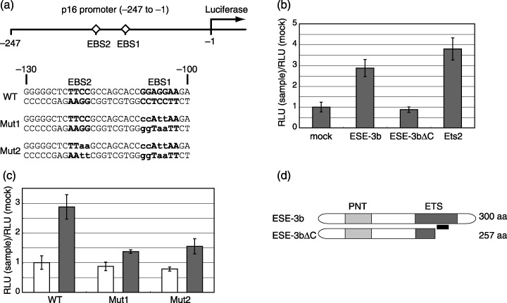
ESE‐3b activates p16INK4a promoter. (a) The p16INK4a promoter construct (–247) was fused with the luciferase gene.( 10 ) Potential Ets‐binding sequences (EBS1 and EBS2) present in the region are indicated by bold letters. Sequences of mutant –247 reporter constructs (Mut1 and Mut2( 10 )) are shown with substituted nucleotides in lowercase. Nucleotide numbers are counted from the translation initiation site. (b) Reporter assays of wild‐type –247 construct in young WI‐38 cells. Empty pcDNA3 (mock), pcDNA3 encoding the wild‐type ESE‐3b (ESE‐3b), the C‐terminally truncated form of ESE‐3b (Ese‐3bΔC), or Ets2 was cotransfected with the –247 reporter plasmid into young WI‐38 cells. Reporter activity was calibrated to the transfection efficiency. The value obtained for the mock experiment was set at 1. RLU, relative luciferase unit. (c) Reporter assays of the wild‐type (WT), Mut1 and Mut2–247 constructs in young WI‐38 cells. Empty pcDNA3 (white bars) or HA‐ESE‐3b‐expressing pcDNA3 was cotransfected with the indicated reporter plasmids into young WI‐38 cells. Reporter activity was calibrated to transfection efficiency, and activity measured by the indicated reporters is shown. (d) Schematic of ESE‐3b (wild‐type) and ESE‐3bΔC (C‐terminal truncated mutant) is shown. Light and dark gray boxes represent PNT (pointed) and Ets domains, respectively. The PNT domain is suggested to function as a protein–protein interaction domain. Black bar indicates a potential nucleotide‐associating site within the Ets domain.