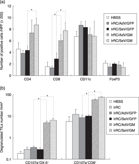
Figure 8.

Immunophenotypic analyses of tumor‐infiltrating leukocytes by immunohistochemistry and flow cytometry. (a) RENCA‐bearing mice were either left untreated (HBSS) or treated with indicated tumor vaccine cells (irRC/AdV/GFP, irRC/SeV/GFP, irRC/AdV/G, and irRC/SeV/G). Resected RENCA tumors were then subjected to immunohistochemical evaluation. To evaluate the distribution of CD4+ T, CD8+ T, CD11c+, and FoxP3+ cells in tumors, positively stained cells were enumerated microscopically at ×200 magnification in 30–70 high‐power fields. (b) Enriched viable lymphocytes from mice treated with the tumor vaccination indicated were stained with anti‐CD8, anti‐DX‐5, and antimouse CD107a antibodies and then subjected to flow cytometry. The cell density (divided by the indicated tumor volume [mm3]) of natural killer cells or CD8+ T cells coexpressing degranulated marker of CD107a (CD107a+NK+ or CD107a+CD8+ T cells) in tumor‐infiltrating leukocytes is shown. Bar graphs depict the means ± SEM. Significant differences are denoted with asterisks (*P < 0.05).
