
Figure 2.
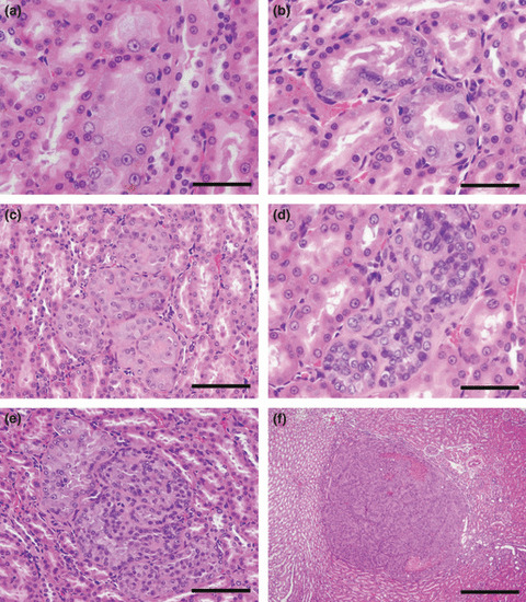
Representative preneoplastic/neoplastic lesions observed in the outer medulla of kidneys. (a,b) Atypical tubules are normal in size, but consist of epithelial cells showing atypia. Bar = 50 μm. (c,d) Atypical hyperplasias are composed of up to 10 tubules containing single‐ or multilayered atypical cells. Bars = 100 μm and 50 μm for (c) and (d), respectively. (e) A renal cell adenoma compressing surrounding tissue. Bar = 100 μm. (f) A renal cell carcinoma with focal necrosis. Bar = 500 μm.