
Figure 1.
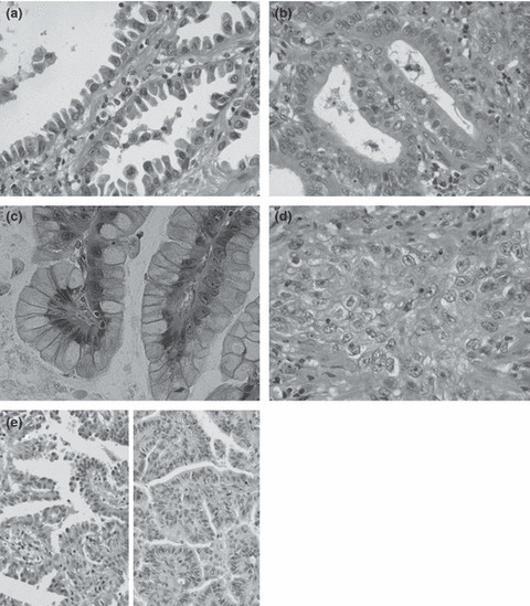
Cell types of adenocarcinomas. (a) Hobnail cell type: epithelial cells look like Clara or type II pneumocyte cells. Apical portions protrude or bulge into the lumen. Note hobnail‐ or tadpole‐shaped cells. (b) Columnar/cuboidal (col/cub) cell type: characterized by rather large columnar or cuboidal cells with flat apices, resembling ciliated cells of bronchial epithelium; cytoplasmic mucus is usually absent, and even when present, is scanty and located near the free cell surface. (c) Goblet cell type: cells have abundant mucus in the cytoplasm, very similar to goblet cells. (d) Polygonal/oval (po/ov) cell type: composed of polygonal or oval cells with or without mucus in the cytoplasm, proliferating in sheets or nests. (e) Mixed cell type: showing a mixture of hobnail (left) and col/cub cells (right) forming a papillary structure. This type usually consists of two from types (a) to (c). Hematoxylin–eosin staining; original magnification: (a–d) ×400; (e) ×200.