
Figure 2.
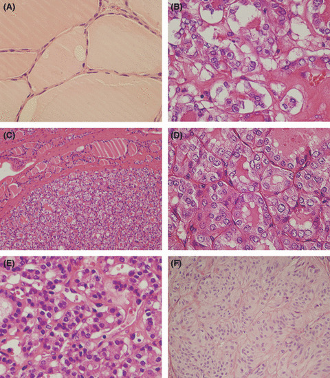
Comparison of morphology in different follicular thyroid lesions. (A) Normal thyroid epithelium. (B) Follicular variant papillary thyroid carcinoma, which shows typical nuclear changes of papillary thyroid carcinoma. (C,D) Well‐differentiated tumor of uncertain behavior, which shows 2–4‐times enlargement of nuclei size, ground glass features, and 3–4% of nuclear grooves, but pseudoinclusions are rare or absent. (E) Follicular thyroid adenoma, which shows 1–4‐times enlargement of nuclei size, the obvious difference is that the nuclei are hyperchromatic. (F) Hyalinizing trabecular adenoma, which is encapsulated, with trabecular growth pattern, polygonal and elongated cells, pseudoinclusions, and cytoplasmic yellow bodies. There is an accumulated hyaline substance in the basement membrane between trabeculae and alveoli. Original magnification: (C,F) ×100; (A,B,D,E) ×200.