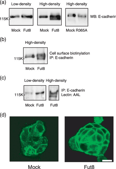
Figure 2.

Effect of α1,6‐fucosyltransferase (Fut8) transfection on the characteristics and the expression of E‐cadherin in colon carcinoma WiDr cells. (a) Western blotting analysis of E‐cadherin in mock, Fut8, and R365A mutated Fut8 transfectants. A whole cell lysate (20 µg) was prepared from low (~3 × 10⁴ cells/cm2) or high‐density (~11 × 104 cells/cm2) cultures and subjected to 8% sodium dodecyl sulfate–polyacrylamide gel electrophoresis (SDS‐PAGE), transferred to a nitrocellulose membrane, and E‐cadherin was detected using an anti‐E‐cadherin antibody. WB, Western blotting. (b) Cell surface expression of E‐cadherin in the mock and Fut8 transfectants. E‐cadherin was immunoprecipitated from whole cell lysate of surface biotinylated mock and Fut8 transfectants, and subjected to 8% SDS‐PAGE, transferred to nitrocellulose membranes and the biotinylated E‐cadherin was visualized using a Vectastain ABC kit and an enhanced chemiluminescence kit. (c) Lectin blot analysis of E‐cadherin in the mock and Fut8 transfectants. E‐cadherin was immunoprecipitated from 400 µg of whole cell lysate, subjected to 8% SDS‐PAGE, and transferred to nitrocellulose membranes, which were probed by Aleuria aurantia lectin (AAL). IP, immunoprecipitation. (d) Distribution of E‐cadherin in mock and Fut8 transfectants. E‐cadherin was detected by a laser scanning confocal microscopy using an anti‐E‐cadherin antibody, ECCD‐2 (scale bar, 10 µm).
