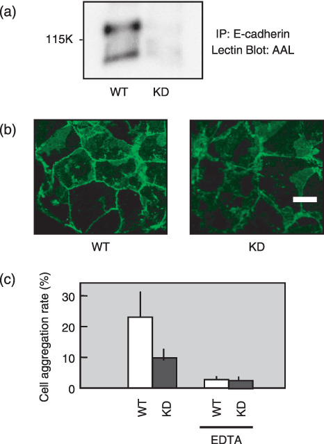
Figure 4.

Changes of E‐cadherin expression and E‐cadherin‐dependent cell–cell adhesion in α1,6‐fucosyltransferase (Fut8) knock down cells. (a) Lectin blot analysis of E‐cadherin in Fut8 knocked down TGP49 cells and wild‐type TGP49 cells. E‐cadherin was immunoprecipitated from 400 µg of whole cell lysate, subjected to 8% sodium dodecyl sulfate–polyacrylamide gel electrophoresis (SDS‐PAGE), and transferred to nitrocellulose membranes, which were probed by Aleuria aurantia lectin (AAL). IP, immunoprecipitation. (b) Distribution of E‐cadherin in Fut8 knocked down TGP49 cells and wild‐type TGP49 cells. E‐cadherin was detected by a laser scanning confocal microscopy using an anti‐E‐cadherin antibody, ECCD‐2. WT, wild‐type TGP49 cells; KD, Fut8 knocked down TGP49 cells (scale bar, 10 µm). (c) Cell–cell aggregation was assayed with or without EDTA. Data represent the mean (±SD) of six experiments.
