
Figure 1.
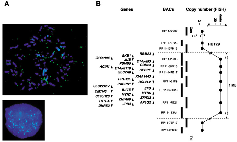
(A) Fluorescence in situ hybridization (FISH) images with RP11‐68M15 as a probe (green signals) hybridized to metaphase (upper) and interphase (lower) chromosomes from the non‐small cell lung cancer line HUT29 clearly showed amplification with a double minute chromosome (dmin) pattern. (B) Map of 14q11.2 covering the region of amplifications observed in the HUT29 cell line. Bacterial artificial chromosomes used for FISH are indicated as thick vertical bars. Within the amplicon, 28 genes exist and are indicated by arrowheads showing the direction of transcription (see Supplementary Table S2).