Figure 3.

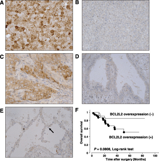
Immunohistochemical staining of BCL2L2 protein. (A) HUT29 cells used as a positive control. (B) A549 cells used as a negative control. (C) Representative case of primary lung adenocarcinoma showing BCL2L2 overexpression compared with normal type II alveolar cells (×400). (D) Representative case of primary lung adenocarcinoma without BCL2L2 overexpression compared with normal type II alveolar cells (×400). (E) Normal primary lung epithelial cells. Black arrowhead indicates the type II alveolar cell. (F) Kaplan–Meier analyses and log‐rank tests in 61 primary lung adenocarcinomas demonstrated that cases with BCL2L2 overexpression tended to be associated with a poorer overall survival compared with those without BCL2L2 overexpression, although the difference was not significant (P = 0.0808).
