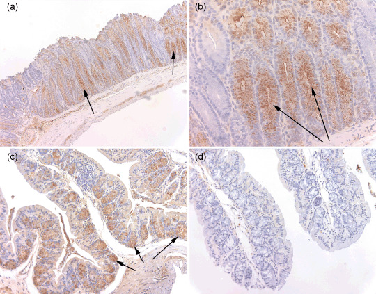
Figure 2.

Immunohistochemistry for Pla2g2a in colon of Muc2−/– Pla2g2a transgenic mice. Expression patterns of Pla2g2a protein were determined by immunohistochemistry using a polyclonal rabbit antimouse antibody at 1:90 000 dilution and visualized with avidin–biotin complex reagents. Formaldehyde‐fixed tissues were obtained from the duodenum, jejunum and ileum and several regions of the colon. In the small intestine Pla2g2a was expressed normally, with strong staining seen at the bottom of crypts in the Paneth cell compartment (data not shown). Figure 2 depicts a representative tissue section from the large intestine. (a) Abundant secreted Pla2g2a protein found in the columnar epithelial cells and lumen of crypts is clearly lacking in cells of goblet cell morphology (200×). Only columnar epithelial cells can be seen lining the crypt. Arrows point to regions of Pla2g2a expression. (b) A higher resolution image of Pla2g2a staining in C57BL/6J‐Muc2−/– Plag2g2a transgenic mice (400×). Arrows point to Pla2g2a expression. (c) Pla2g2a immunostaining from tissue from a C57BL/6J Pla2g2a transgenic mouse that is wildtype for Muc2 (40×). Pla2g2a is clearly detected in the goblet cells but not the enterocytes at the crypt borders. (d) Immunostained tissue from a C57BL/6J mouse (200×). No Pla2g2a protein is detected.
