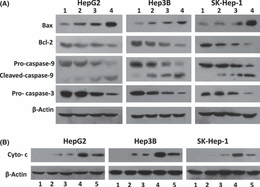
Figure 5.

Expression of apoptosis‐related proteins in vitro. (A) HepG2, Hep3B, and SK‐Hep‐1 cells were treated with 2 μm arsenic trioxide (ATO) (lane 2), genistein (GEN) (15 μm for HepG2 and SK‐Hep‐1, 20 μm for Hep3B) (lane 3), or ATO + GEN (lane 4), for 48 h. Untreated cells served as the control (lane 1). The cells were homogenized and subjected to Western blot analysis to detect the expression of Bax, Bcl‐2, pro‐caspase‐9, cleaved caspase‐9, and pro‐caspase‐3. β‐Actin served as an internal control. (B) Cytoplasmic proteins from each of the treated cells as in (A) (lanes 1 to 4) were prepared using a Mitochondria/cytosol Fractionation Kit. As an additional control, cytoplasmic proteins were prepared from cells pretreated with N‐acetyl‐l‐cysteine (NAC) followed by ATO + GEN (lane 5). The expression of cytochrome c (Cyto‐c) in the mitochondria‐depleted cytosolic fractions was examined by Western blot analysis. β‐Actin served as an internal control.
