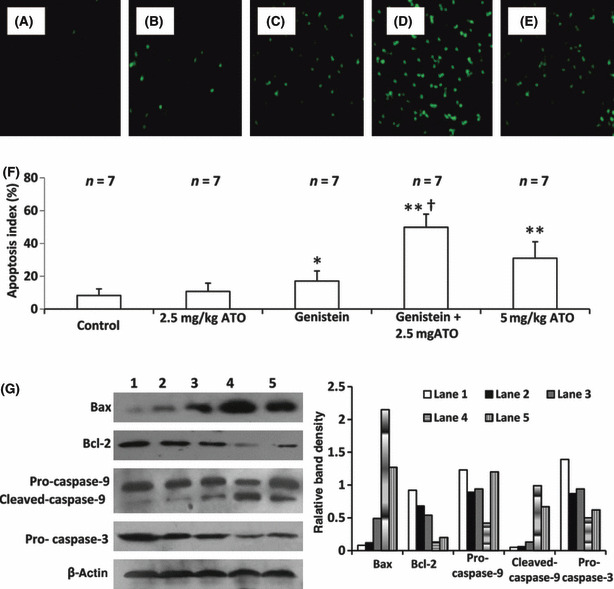
Figure 8.

Genistein (GEN) synergizes with arsenic trioxide (ATO) to induce cell apoptosis in situ. Illustrated are representative tumor sections prepared from mice that received daily injections of PBS (control) (A), 2.5 mg/kg ATO (B), genistein (C), genistein + 2.5 mg/kg ATO (D), or 5 mg/kg ATO (E) as in Fig. 6. The sections were stained with the TUNEL agent to visualize apoptotic cells. (F) TUNEL‐positive cells were counted to calculate the apoptosis index. n, number of tumors assessed. *Significant difference in the apoptosis index from control; **highly significant difference at P < 0.001 from control; †significant difference from ATO monotherapy. (G) The tumor tissues were homogenized and subjected to Western blot analysis to detect expression of Bax, Bcl‐2, pro‐caspase‐9, cleaved caspase‐9, and pro‐caspase‐3 (left panel). The numbering of lanes is as in Figure 5(b). β‐Actin served as an internal control. The band density was measured and compared to that of β‐actin to calculate relative band density (right panel).
