
Figure 3.
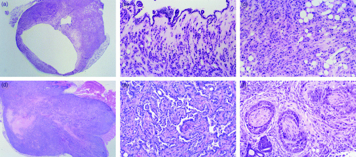
(a) MGC1 cells transplanted subcutaneously into nude mice grew slowly to form cystic tumors, consisting of a monolayer of rather large, pleomorphic, hyperchromatic cells. (× 10 H&E). (b) High‐power view of Fig. 3a. Cystic tumor formation with monolayer of MGC1 cells. (× 40 H&E). (c) High‐power view of Fig. 3a. Tumor formation with MGC1 cells. (× 40 H&E). (d) MGC2 cells transplanted subcutaneously into nude mice grew with stable transplantation and glandular‐like configurations. The tumors were composed of rather small, pleomorphic, hyperchromatic cells arranged in a solid pattern. (× 10 H&E). (e) High‐power view of Fig. 3d. Tumor tended to form glandular‐like constructs with MGC2 cells. (× 40 H&E). (f) Transplanted MGC2 cells with perineural invasion. MGC2 cells grew invasively under the transplanted tissue, with perineural invasion and invasion to the muscle layer. (× 40 H&E).