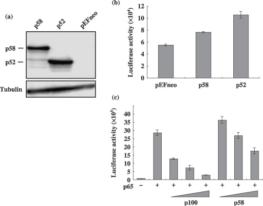
Figure 7.

Transcriptional activity of NF‐κB2/p58 in a T‐cell line. (a) Cell lysates were prepared from 293T cells transfected with either the pEFneo‐p58, pEFneo‐p100 or pEFneo plasmid (2 µg), and the amount of NF‐κB2 protein in each lysate was measured by a Western blot analysis using an antip100 antibody. The arrows indicate the p58 and p52 recognized by the antibody. (b, c) Jurkat cells were transfected with either the pEFneo‐p58, pEFneo‐p100 or pEFneo plasmid (0.1 µg) together with the luciferase plasmid (0.5 µg) regulated by the NF‐κB element (kB‐Luc) and the β‐galactosidase plasmid (0.1 µg). The pSG‐p65 plasmid (0.1 µg) was cotransfected with increasing amount of pEFneo‐p100 or pEFneo‐p58 (0.05, 0.1, and 0.2 µg) into Jurkat cells as indicated in (c). Cell lysates were prepared from transfected cells, and the luciferase and β‐galactosidase activities were determined. The luciferase activity normalized by the β‐galactosidase activity was shown as the average with standard deviations. Three independent experiments were carried out to confirm reproducibility.
