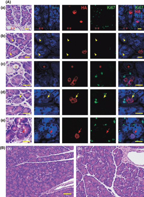
Figure 2.

Acinar cell‐specific expression of hemagglutinin (HA)‐KrasG12V. (A) Localization of amylase (blue) protein, HA‐KrasG12V (red) and Ki67 (green) at 2 days after injection of virus with Ad‐Amy‐Cre (a, b, c, d) and Ela‐Cre (e). All the HA‐KrasG12V positive cells (red) were acinar cells; expression was not observed in duct cells (**), centroacinar cells (yellow arrowhead), or small duct cells (*). Most virally infected acinar cells positive for HA‐KrasG12V were indistinguishable from non‐infected acinar cells by hematoxylin–eosin staining. Some infected acinar cells have nuclei with a so‐called “owl‐eye” (yellow arrows) or “ground glass” (red arrows) appearance. Ki67 (green) is not present in the nuclei of the cells expressing HA‐KrasG12V (red). Bar, 20 μm. (B) None of the Ad‐Amy‐Cre (a) or the Ad‐Ela‐Cre (b) groups displayed any pancreatic lesions, even after 8 weeks. Bar, 50 μm.
