
Figure 4.
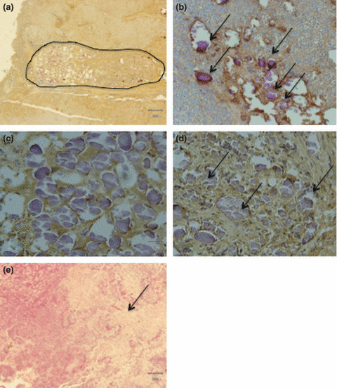
Histopathologic findings of the FU‐MMT‐3 xenografts. A feeding artery (encircled) was entirely embolized by β‐tricalcium phosphate (TCP) ceramic microspheres. (a) Fibrosis with granuloma formation and lymphocytic infiltration was observed in the lumen of the feeding arteries; no injury caused by microspheres was found in the vessel walls (×40). (b) TCP microspheres were remarkably embolized (arrows) in various‐sized microvessels (×200). (c) TCP microspheres showing uniform basophilia (×400). (d) Aggregates of TCP microspheres (arrows) in tumor microvessels (×400). (e) Coagulative necrosis (arrows) is extensively seen in treatments with microspheres loaded with or without TNP‐470 (×40).