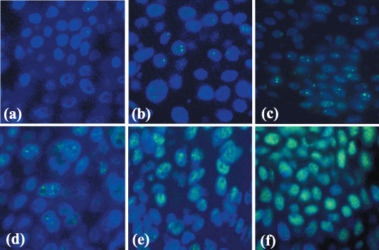
Figure 2.

Immunofluorescence of p53‐binding protein 1 (53BP1) expression in human skin tumors. (a) Seborrheic keratosis (SK) in the non‐exposed skin expressed stable type staining with rarely one nuclear focus (stable type), (b) whereas SK in the sun‐exposed skin expressed an occasional one or two nuclear foci (low DNA damage response (DRR) type). (c) Actinic keratosis showed three or more discrete nuclear foci in dysplastic cells (high DRR type). (d) Bowen's disease showed several discrete nuclear foci mixed with intense and heterogeneous nuclear staining (mixed DRR and abnormal type). (e) Squamous cell carcinoma as well as (f) basal cell carcinoma exhibited intense and heterogeneous nuclear staining (abnormal type).
