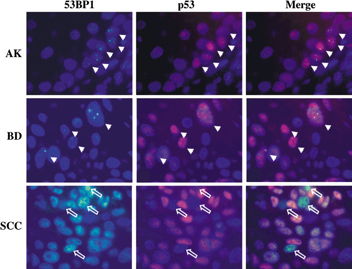
Figure 3.

Double‐labeled immunofluorescence for p53‐binding protein 1 (53BP1) and p53 expression. Actinic keratosis (AK) showed colocalization of discrete 53BP1 nuclear foci and p53 nuclear staining in dysplastic cells at the basal layer, suggesting an activation of DNA damage response (DDR). Bowen's disease (BD) also showed colocalization of discrete 53BP1 nuclear foci and p53 nuclear staining in dysplastic cells including dispersed plump cells. Squamous cell carcinoma (SCC) exhibited intense and heterogeneous nuclear staining of both 53BP1 and p53 immunoreactivity; however, intense 53BP1 staining was not always colocalized with p53 overexpression, suggesting a disruption of the DDR pathway. Arrows indicate colocalization of 53BP1 nuclear foci and p53 staining in both AK and BD. Open arrows indicate cancer cells showing intense 53BP1 staining with no p53 staining in SCC.
