
Figure 1.
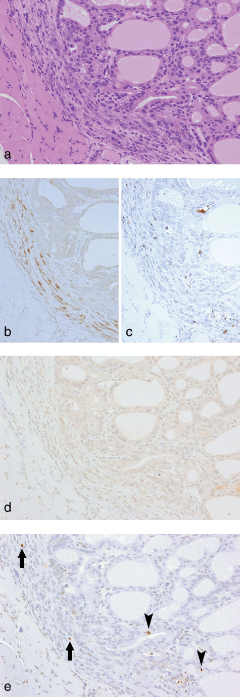
Slight inflammatory cell infiltration with focal migration of epithelial cells from disrupted follicles into the thyroid capsular region at week 4 in a rat treated with sulfadimethoxine following N‐bis(2‐hydroxypropyl)nitrosamine‐initiation. (a) H&E. Original magnification ×180. (b) A serial section of (a). CD3 immunohistochemistry. Some inflammatory cells are positive for CD3, indicative of a T‐cell nature. (c) A serial section of (a). ED1 immunohistochemistry. A few inflammatory cells are positive for ED1, indicative of a macrophage nature. (d) A serial section of (a). inducible nitric oxide synthase immunohistochemistry. No positive cells were found. (e) A serial section of (a). Single‐strand DNA (ssDNA) immunohistochemistry. A few ssDNA‐positive dot signals, demonstrating fragmented nuclei, are evident in some inflammatory and/or interstitial cells (arrows) and follicular epithelial cells (arrowheads).