
Figure 1.
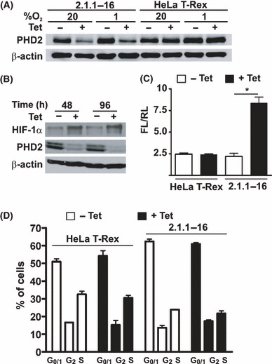
Characterization of the 2.1.1‐16 tetracycline (Tet)‐inducible prolyl‐4‐hydroxylase domain 2 (PHD2) knockdown cell line. (A) HeLa T‐Rex and 2.1.1‐16 cells were incubated in the presence or absence of 10 μg/mL Tet for 96 h at 20% O2 or 1% O2. Subsequently, cells were lysed and protein extracts were analyzed by immunoblot. (B) The 2.1.1‐16 cells were incubated for 48 or 96 h in the presence or absence of 10 μg/mL Tet at 20% O2. Subsequently, cells were lysed and protein extracts were analyzed by immunoblot. (C) HeLa T‐Rex and 2.1.1‐16 cells were incubated in the presence or absence of 10 μg/mL Tet for 48 h. Subsequently, cells were transfected with the firefly luciferase (FL) hypoxia‐inducible factor (HIF) reporter gene plasmid pH3SVL and the renilla luciferase (RL) control plasmid pRLSV40. After 24 h transfection cells were lysed and FL and RL activities were determined. (n = 3 ± SEM) *P < 0.05. (D) HeLa T‐Rex and 2.1.1‐16 cells were incubated in the presence or absence of 10 μg/mL Tet for 48 h. Subsequently, cell cycle distribution was analyzed by FACS (n = 3 ± SD).