
Figure 3.
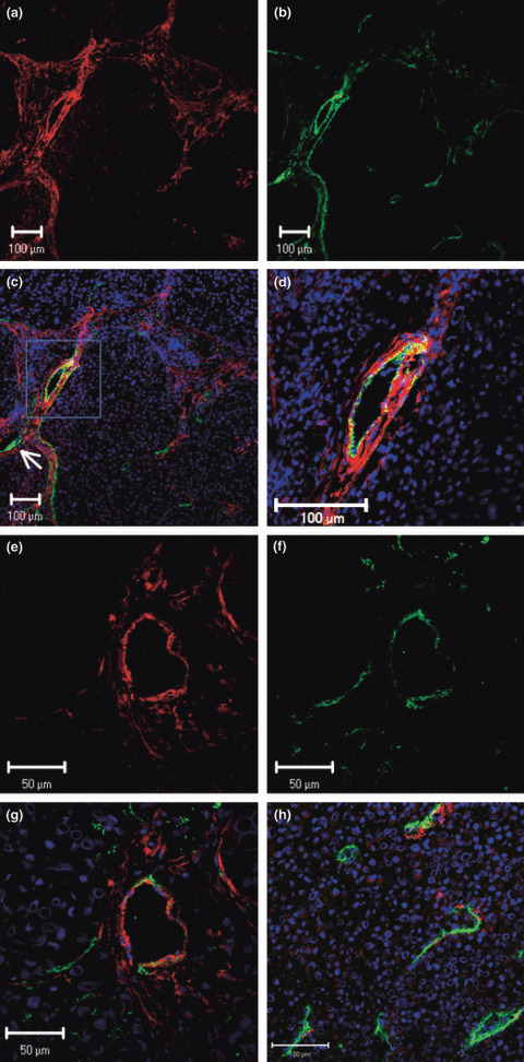
Fluorescence double‐labeled immunohisto‐chemistry (IHC) of TMK‐1 human gastric cancer cells growing in nude mice. Representative images show IHC for PDGF‐Rβ in red and Lyve‐1 (lymphatic endothelial marker) in green. (a–g) PDGF‐Rβ was detected in lymphatic endothelial cells on enlarged and tortuous lymphatic vessels located immediately adjacent to tumor nests. Small lymphatic vessels (arrow) did not express PDGF‐Rβ. (h) Intratumoral lymphatic vessels did not express PDGF‐Rβ.