
Figure 1.
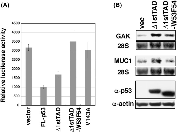
Δ1stTAD‐inducible genes in Δ1stTAD‐dependent apoptosis. (A) Luciferase gene cotransfection growth analysis was performed. Saos2 cells (2 × 10⁴ cells) were transfected with 15 ng of pcDNA3, pcDNA3‐FL‐p53, pcDNA3‐Δ1stTAD, pcDNA3‐Δ1stTAD‐W53F54 or pcDNA3‐V143A together with 2 ng of PicaGene control vector. Cells were harvested 48 h post‐transfection, and luciferase activities were measured. (B) Expressions of GAK and MUC1 were analyzed by Northern blotting. Saos2 cells (5 × 10⁶ cells) were transfected with 3.75 µg of pcDNA3, pcDNA3‐Δ1stTAD or pcDNA3‐Δ1stTAD‐W53F54. Cells were harvested 24 h post‐transfection and analyzed by Northern blotting as in Fig. 3B. Expression of each p53 was analyzed by Western blotting (bottom panel).