Figure 2.

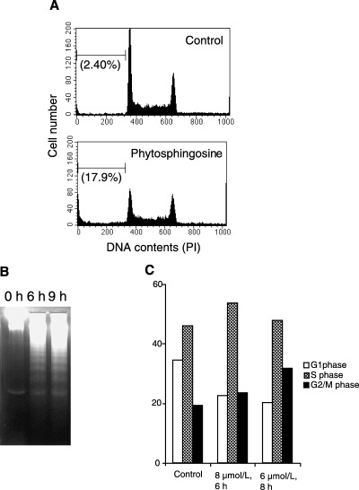
Phytosphingosine‐induced apoptosis and cell cycle arrest in Jurkat cells. (a) Jurkat (neo) cells were incubated with 0 or 8 µM phytosphingosine for 6 h. At the indicated times, the cells were collected and permeabilized. Cells were then stained with PI, and the DNA contents were determined by flow cytometry. Values in parentheses indicate sub‐G1 cell percentages. Data are representative of three independent experiments. (b) Jurkat (neo) cells were incubated with 8 µM phytosphingosine for 0, 6, and 9 h. At the times indicated, the cells were lyzed and DNA was prepared. DNA fragmentation was analyzed by agarose gel electrophoresis. (c) Jurkat (neo) cells were incubated with or without phytosphingosine for indicated times and doses. Percentage of cells in each cell cycle phase were measured. Data are representative of three independent experiments.
