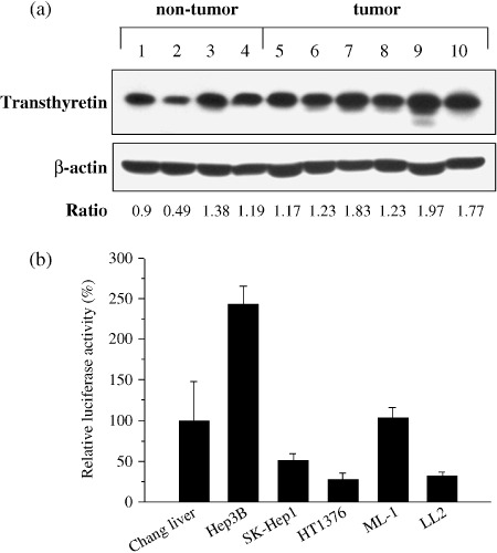
Figure 1.

Detections of transthyretin protein and transcriptional activity of transthyretin promoter in various human and murine cells. (a) Transthyretin protein was produced in the non‐tumor (Lanes 1–4) and tumor (Lanes 5–10) parts of the liver from hepatocellular carcinoma (HCC) patients, as determined by immunoblot analysis. Lanes 1 and 6, 3 and 9, and 4 and 7 were paired samples. Expression of β‐actin served as the quantitative control. P = 0.04 for tumor group vs. non‐tumor group by unpaired Student's t‐test. (b) The activity of the transthyretin promoter was higher in liver cell lines compared with non‐liver cell lines. Cells were transfected with pGL‐TTRp and the transfection efficiency was standardized to the cotransfected plasmid pTCYLacZ expressing β‐galatosidase driven by the β‐actin promoter. Relative luciferase activity was indicated by comparing each cell line with Chang liver cells. Each value represents the mean ± SD (n = 3).
