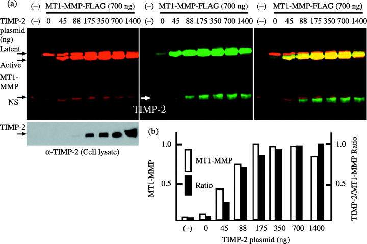
Figure 1.

Cell‐surface membrane‐type (MT) 1‐matrix metalloproteinase (MMP) and tissue inhibitor of metalloproteinase‐2. (a) The expression plasmid for MT1‐MMP‐FLAG (700 ng) was cotransfected with the indicated amounts of tissue inhibitor of metalloproteinase‐2 plasmid into 293T cells. At 48 h after transfection, cells were labeled with biotin, and were analyzed as described in ‘Materials and Methods’. Cell‐surface MT1‐MMP and tissue inhibitor of metalloproteinase‐2 (TIMP‐2) coprecipitated with MT1‐MMP were detected with IRDye 800‐conjugated streptavidin (middle panel), whereas MT1‐MMP latent and active forms were both detected with anti‐FLAG M2 antibody labeled with Alexa Fluor 680 (left upper panel). Aliquots of cell lysates were examined for the expression of TIMP‐2 by western blotting using anti‐TIMP‐2 antibody (left lower panel). Merged 800 and 680 signals are shown in the right panel. NS, non‐specific band. (b) Cell‐surface MT1‐MMP levels and the ratio of TIMP‐2 to MT1‐MMP signal measured from the middle panel are shown. The highest MT1‐MMP level (175 ng) and TIMP‐2/MT1‐MMP ratio (1400 ng) were arbitrarily set to 1, and the levels of other transfections were adjusted accordingly. The data represent one of three different experiments showing similar results.
