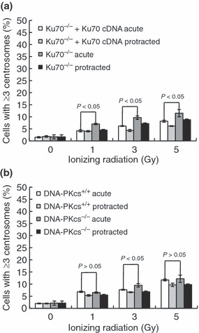
Figure 4.

Centrosome overduplication in non‐homologous end‐joining repair protein‐deficient cells. (a) Ku70−/− fibroblast cells and complemented Ku70−/−+Ku70 cDNA cells were stained with anti‐γ‐tubulin antibodies and anti‐centrin‐2 antibodies after irradiation with 5 Gy at dose rates of 1 Gy/min and 0.5 mGy/min. Cells were irradiated with the indicated doses and centrosomes were quantified 72 h after irradiation. *Student’s t‐test indicated a statistically significant increase (P > 0.05). (b) As in (a), DNA‐PKcs−/− fibroblast cells (SCID) and parental CB17 mouse fibroblast cells (DNA‐PKcs+/+) were stained, and centrosomes were quantified 72 h after irradiation. Bars represent the standard errors (n = 3). *Student’s t‐test indicated no statistically significant increase (P < 0.05).
