Figure 2.

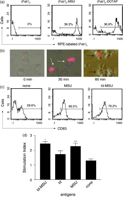
Monosodium urate (MSU) crystals affected dendritic cells (DC). (a) Phagocytotic ability of DC pulsed with R‐phycoerythrin (RPE)‐labeled (Fab′)2‐MSU complex ([Fab′]2‐MSU), RPE‐labeled (Fab′)2 conjugated with N‐(2,3‐dioleoyloxy‐1‐propyl)trimethylammonium methyl sulfate ([Fab′]2‐DOTAP) or RPE‐labeled (Fab′)2 ([Fab′]2) after 1 h incubation was examined by flow cytometry. Results are representative of two similar independent experiments. (b) Engulfment of RPE‐labeled (Fab′)2 attached to MSU crystals was evaluated by fluorescence microscopy. The white arrow shows RPE‐labeled (Fab′)2‐MSU complex attached to DC, while the black arrow shows RPE‐labeled (Fab′)2 was incorporated into DC under fluorescence microscopy. Scale lines, 20 µm. (c) CD83 expression in DC pulsed with MSU crystals, idiotype (Id)‐MSU complex or none for 48 h was examined by flow cytometry. Results are representative of at least two similar experiments. (d) T‐cell proliferation activity of DC pulsed with Id‐MSU complex. Autologous CD3+ cells were co‐cultured with a fixed number of DC (T : DC 10:1) for 5 days. Stimulation index (SI) was calculated as the ratio of formazan levels in the sample over background formazan levels. *P < 0.01 (vs Id alone) **P < 0.01 (vs none). Data are expressed as mean ± standard deviation of three volunteers in triplicate.
