
Figure 1.
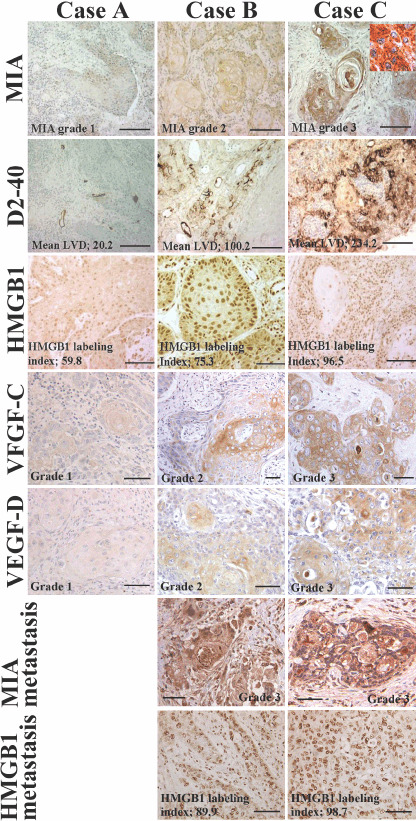
Immunohistochemical staining of melanoma inhibitory activity (MIA), D2‐40, high mobility group box‐1 (HMGB1), vascular endothelial growth factor (VEGF)‐C and VEGF‐D in oral squamous cell carcinomas (OSCC). The expressions of D2‐40, HMGB1, VEGF‐C and VEGF‐D were shown in case A (T2N0M0, stage II, well differentiated OSCC), case B (T2N1M0, stage III, well differentiated OSCC) and case C (T4N2M0, stage IV, well differentiated OSCC). Inset showed MIA localization at the cytoplasm and cytoplasmic membrane. Expressions of MIA and HMGB1 were examined in lymph node metastasis in case B and C. Bar, 100 µm.