
Figure 5.
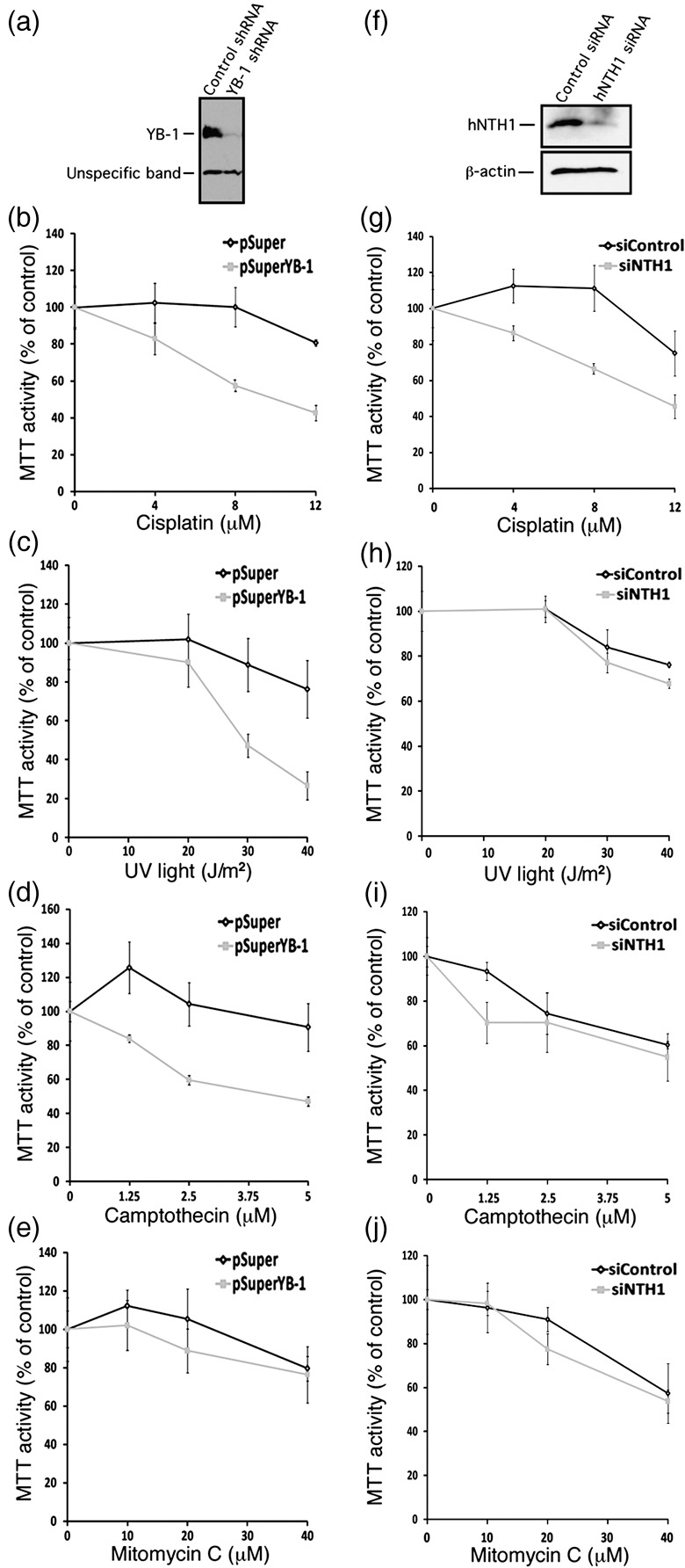
Impact of depleting either endogenous Y‐box‐binding protein‐1 (YB‐1) or human endonuclease III (hNTH1) proteins in MCF7 on survival after different DNA‐damaging conditions. (a) Western blots showing intracellular levels of YB‐1 48 h after transfection with a shRNA specific to YB‐1 or with a control construct. The lower molecular weight band is an unspecific protein recognized by the anti‐YB‐1 antibody. (b–e) MTT cell survival assays of MCF7 cells transfected with a shRNA specific to YB‐1 or with a control construct after of treatment with either cisplatin (b), UV light (c), camptothecin (d), or mitomycin C (e). Cells were first transfected with the appropriate constructs. Forty‐eight hours later, cells were plated in 96‐well plates. Cells were then treated with the drugs for 16 h in culture before the MTT assays. Results represent the mean ± SD of triplicate experiments. (f) Western blots showing intracellular levels of hNTH1 after 48 h after transfection with a siRNA specific to hNTH1 or with a scrambled control siRNA. The anti β‐actin was used as loading control for the blot. (g–j) MTT cell survival assays of MCF7 cells transfected with a siRNA specific to hNTH1 or with a scrambled control siRNA after treatment with either (g) cisplatin, (h) UV light, (i) camptothecin, or (j) mitomycin C. Cells were first transfected with the siRNAs. Forty‐eight hours later, cells were plated in 96‐well plates. Cells were then treated with the drugs for 16 h in culture before the MTT assays. Results represent the mean ± SD of triplicate experiments.