
Figure 2.
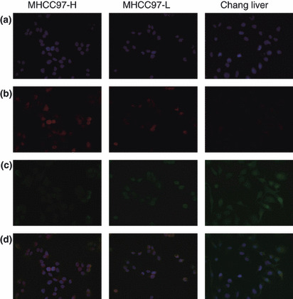
Double staining of osteopontin (OPN) and caspase‐3 by immunofluorescence: (a) DAPI staining. (b) OPN was highly enriched in the cytoplasm of MHCC97‐H cells, moderately so in MHCC97‐L cells, and weakly so in Chang liver cells. (c) Caspase‐3 expression increased as metastatic potential decreased in hepatocellular carcinoma (HCC) cell lines. (d) OPN and caspase‐3 expression in different cell lines (magnification, ×200).