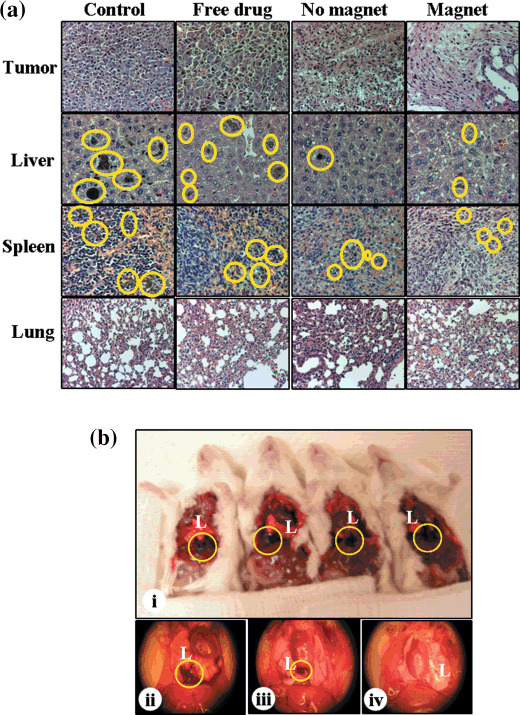
Figure 5.

Histopathology. Formalin‐fixed, paraffin‐embedded tissue sections of different organs such as the tumor, liver, spleen, and lung were stained with hematoxylin–eosin (a). Magnification 20× (bar = 10 µm). Pictures of tumor metastasis of pleural cavity (b). Control mice (b[i]), free vinblastine (b[ii]), vinblastine‐loaded magnetic cationic liposomes (MCLs) without magnet (b[iii]) and with magnet (b[iv]). Pictures are representative of mice from each treatment group. The circle denotes melanin pigment, reveals invasiveness of melanoma tumor in liver, spleen, and pleural cavity.
