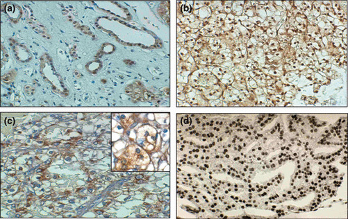
Figure 1.

Examples of p27kip1 immunohistochemical staining in renal epithelial tumors. (a) Normal renal tissue showing nuclear staining of tubular epithelial cells. (b) Example of a well differentiated clear‐cell renal cell carcinoma (RCC) displaying a strong staining for p27kip1. (c) Example of a poorly differentiated clear‐cell RCC displaying a negative nuclear staining for p27kip1 with an evident cytoplasmic positivity (d) Example of immunostaining for 8‐hydroxydeoxyguanosine (8‐OHdG) in a representative case of clear cell RCC displaying a positive staining. Original magnifications: (a) ×200; (b) ×150; (c) ×250; (d) ×200; inset in (c) ×800.
