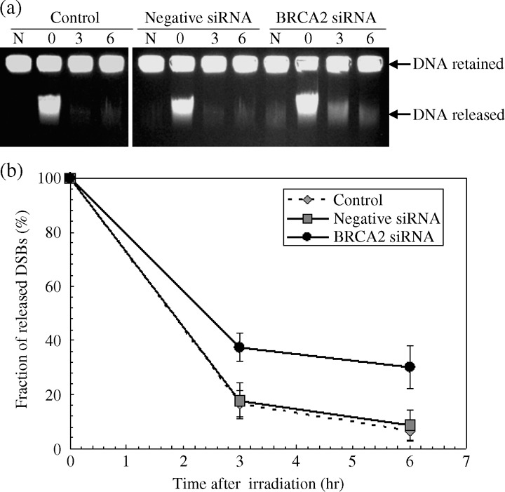
Figure 3.

Rejoining kinetics of radiation induced DNA DSBs in HeLa cells. Cells were transfected with mock‐treated (control), negative siRNA or 20 nM BRCA2 siRNA. 48 h after transfection, cells were irradiated with 20 Gy of X‐rays on ice and incubated for various repair periods at 37°C. (a) Typical ethidium bromide stained gel images by constant‐field gel electrophoresis. N designates non‐irradiated control samples. (b) The fraction of migrated broken DSBs obtained by quantifying the intensity of the bands in gels from (a). Data represent the mean and the standard deviation from three separate experiments, P < 0.05 versus control or mock‐transfected cells (Student's t‐test).
