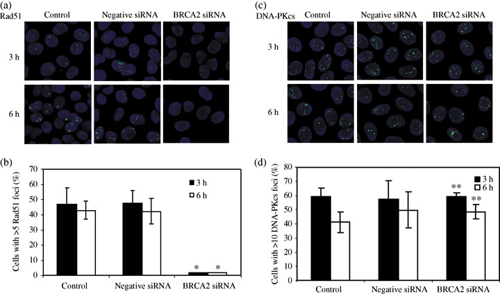
Figure 4.

Foci formations of Rad51 and phosphorylated DNA‐dependent protein kinase catalytic subunit (DNA‐PKcs) in HeLa cells irradiated with 2 Gy X‐rays. Nuclei were stained with diamidino‐2‐phenylindole. The primary antibody was rabbit polyclonal anti‐Rad51 (a) and antiphosphorylated DNA‐PKcs (2609 Thr) (c). Number of cells containing Rad51. (b) and DNA‐PKcs (d) foci visualized at 3 h (filled columns) and 6 h (open columns) after exposure to 2 Gy X‐rays. Data represent the mean and standard deviation from three separate experiments. *P < 0.05 and **P > 0.05 versus control or mock‐transfected cells (Student's t‐test).
