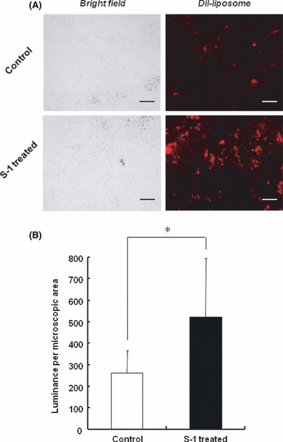
Figure 5.

Effect of S‐1 dosing on intratumoral distribution of polyethylene glycol (PEG)‐coated liposomes. Tumor‐bearing mice, pretreated with S‐1 dosing for 7 days, received DiI (1, 1′‐dioctadecyl‐3, 3, 3′,3′‐tetramethyl‐indocarbocyanine perchlorate)‐labeled PEG‐coated liposomes. At 24 h post‐injection, the section of tumor was examined with fluorescence microscopy. (A) Intratumoral distribution of PEG‐coated liposomes. Red spots represent liposomal distribution. Bar, 100 μm. Original magnification, ×200. (B) Mean fluorescence intensity per microscopic area. Data represent mean ± SD. *P < 0.05.
