
Figure 4.
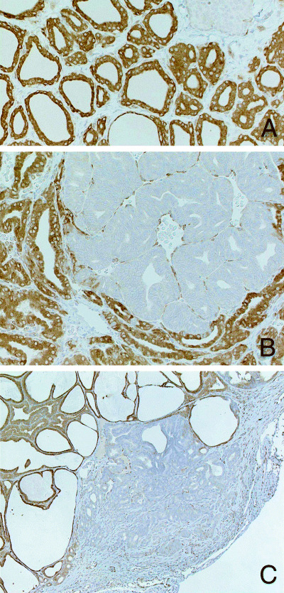
(A) Cytoplasmic positivity for COX‐2 is evident in follicular cells in a control rat. The COX‐2 negative tissue shown on the upper right is parathyroid. Immunohistochemistry (×360). (B) Marked reduction of COX‐2 expression in neoplastic lesions of thyroid follicular cells. Adenoma in a rat treated with PTU for 4 weeks after DHPN‐initiation. Immunohistochemistry (×360). (C) Adenocarcinoma invading into the thyroid capsule in a rat treated with SDM for 10 weeks after DHPN‐initiation. Immunohistochemistry (×140).