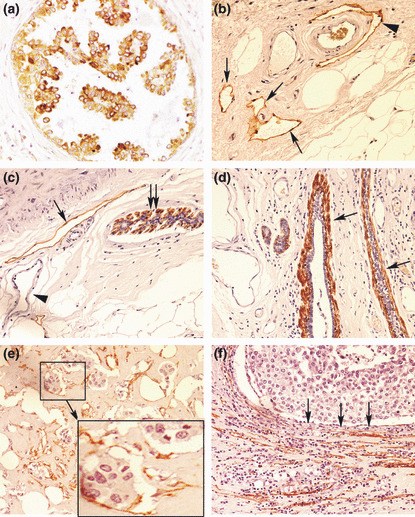
Figure 2.

(a) Vascular endothelial growth factor (VEGF)‐C‐positive cancer cells in ductal carcinoma in situ (DCIS). (b–f) Various types of D2‐40 staining (brown): (b) lymphatics (arrows) in the peritumoral stroma, one of them (arrow head) embracing an artery which is unstained; (c) myo‐epithelial cells in the outermost layer of DCIS (double‐arrows); and a lymphatic (single‐arrow) in the stroma, blood capillaries (arrow head) remaining unstained; (d) myo‐epithelial cells (arrows) comprising the outermost layer of mammary ducts; (e) lymphatics with incomplete endothelial linings containing cancer cells indicative of lymphovascular invasion, shown clearly in the magnified inset of the selected area; (f) collapsed/compressed lymphatics in the stroma, adjacent to lobular cancerous tissue (arrows indicate direction of compression). Original magnifications, ×400.
