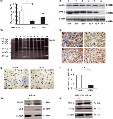
Figure 4.

Effect of mesenchymal stem cells (MSC) on MMP‐2 and MMP‐9 expression in MHCC97‐H cells. Real‐time PCR (A) and western blot analysis (B) showed that both mRNA and protein levels of MMP‐2 in MHCC97‐H were decreased by culture with MSC conditioned medium (MSC‐CM), whereas there was no significant change in the expression of MMP‐9. Lanes 1–3, or 4–6 correspond to 0%, 25% and 50% MSC‐CM, respectively. (C) In zymography analysis, the amount of pro‐MMP‐2 (72 kD) was significantly lower in the MSC‐treated mice (lanes 2, 3, 6, 7) than in the controls (lanes 4, 5, 8–10). (D) Immunohistochemical staining showed lower expression of MMP‐2 in hepatocellular carcinoma (HCC) tissues following MSC treatment (a) compared with the controls (b), whereas no significant alteration in MMP‐9 expression was observed between MSC‐treated animals (c) and controls (d) (×20). (E,F) Transformation growth factor‐beta 1 (TGFβ1) siRNA significantly inhibited the invasive capability of MHCC97‐H cells compared with the controls, as evaluated by Transwell assays. *P < 0.05. (G) Western blot analysis showed that TGFβ1 siRNA concurrently downregulated expression of TGFβ1 and MMP‐2 in MHCC97‐H cells under non‐reducing conditions. (H) The TGFβ1 siRNA significantly blocked the inhibitory effect of MSC on the expression of TGFβ and MMP‐2 in MHCC97‐H cells under non‐reducing conditions.
