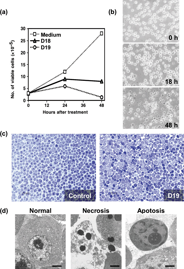
Figure 1.

Effect of mAbs on the proliferation of DT40 cells. (a) DT40 cells were cultured in the presence or absence of monoclonal antibodies (mAbs) (D18 or D19), and cell growth of DT40 cells was evaluated by a trypan blue dye exclusion method. (b) Phase‐contrast micrographs of morphological alteration of DT40 cells cultured with D18 mAb are shown. (c) Semi‐thin sections of DT40 cells cultured for 18 h with or without D19 mAb were stained with toluidine blue solution. (d) Electron microscopic analysis of ultra‐thin sections of DT40 cells cultured for 18 h without (left) or with D19 (middle and right).
