
Figure 2.
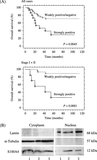
(A) Overall survival of ovarian carcinoma patients according to the nuclear S100A4 expression. Kaplan–Meier analysis shows significantly poorer survival in patients with strong nuclear S100A4 expression compared to those with weakly positive/negative nuclear S100A4 expression. (B) Western blot analysis of cytoplasmic and nuclear fractions obtained from three carcinoma tissues. Lane 1, serous carcinoma shown in Figure 1b; lane 2, endometrioid carcinoma shown in Figure 1C; lane 3, clear cell carcinoma shown in Figure 1D. The presence of S100A4 not only in the cytoplasmic but also in the nuclear fraction was observed in both cases (lanes 2 and 3) that showed nuclear staining of S100A4 (Fig. 1C,D). Lamin B and α‐tubulin were used as the markers for nuclear and cytoplasmic fractions, respectively.