
Figure 3.
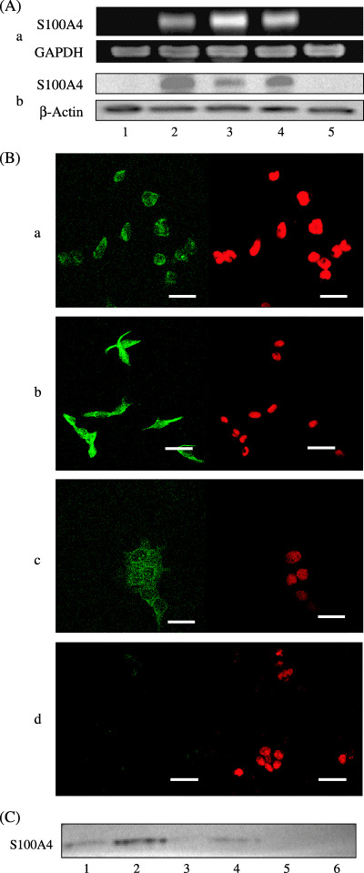
(A) RT‐PCR (a) and Western blot (b) analyses for the expression of S100A4 in normal OSE cells (1) and ovarian carcinoma cells (2, A2780; 3, A2780/CDDP; 4, SKOV3; 5, OVCAR3). S100A4 expressions at both mRNA and protein levels are observed in three ovarian carcinoma cells, but not in normal OSE or ovarian carcinoma OVCAR3 cells. (B) Immunofluorescent analysis for the subcellular localization of S100A4 in ovarian carcinoma cells. A2780 (a) and A2780/CDDP (b) cells show both cytoplasmic and nuclear expressions, whereas SKOV3 cells show only cytoplasmic expression (c) and OVCAR3 cells show faint cytoplasmic expression (d). Left panel, S100A4 staining; right panel, propidium iodide staining. Bar, 40 µm. (C) Western blot analysis for S100A4 in the culture media from ovarian carcinoma cells (lane 1, A2780; lane 2, A2780/CDDP; lane 3, OVCAR3; lane 4, SKOV3) and culture medium without cells (lane 5, RPMI‐1640; lane 6, Dulbecco's modified Eagle's medium). Secretion of S100A4 into the culture media is confirmed in A2780, A2780/CDDP, and SKOV3 cells, but not in OVCAR3 cells.