
Figure 7.
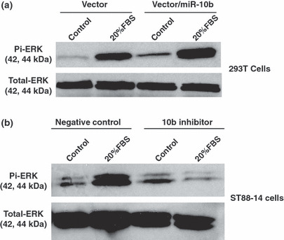
miR‐10b modulated RAS signaling. (a) The MDH1‐PGK‐GFP/miR‐10b (vector/miR‐10b) or MDH1‐PGK‐GFP vector (vector) was transfected into 293T cells. Transfected cells were stimulated with serum free medium (control) or medium supplemented with 20% FBS for 15 min, and phosphorylated ERK and total ERK were analyzed by western blotting. Overexpressing miR‐10b induced higher phosphorylated ERK expression. (b) The miR‐10b inhibitors or negative controls were transfected into NF1 MPNST cells (ST88‐14). Cells were serum‐starved for 24 h, and then incubated in serum‐free medium (control) or medium plus 20% FBS for 15 min. Phosphorylated ERK and total ERK were detected by western blotting. Compared to controls, inhibiting miR‐10b expression reduced phosphorylated ERK in response to serum stimulation.