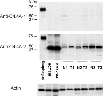
Figure 3.

Western blot analysis of the C4.4A protein in normal esophageal tissue, colorectal cancer cell lines (HCT116 and KM12SM), and in three samples from patients taken from normal (N) and cancerous colorectal tissues (T). Esophageal epithelium displayed an intense band of approximately 67 kDa with both the anti‐C4.4A‐1 and anti‐C4.4A‐2 antibodies. However, in both colon cancer cell lines and colon tissue samples, anti‐C4.4A‐2 antibody alone displayed a prominent band at approximately 40 kDa. Anti‐C4.4A‐1 antibody did not produce any bands. The blot for actin served as a loading control.
