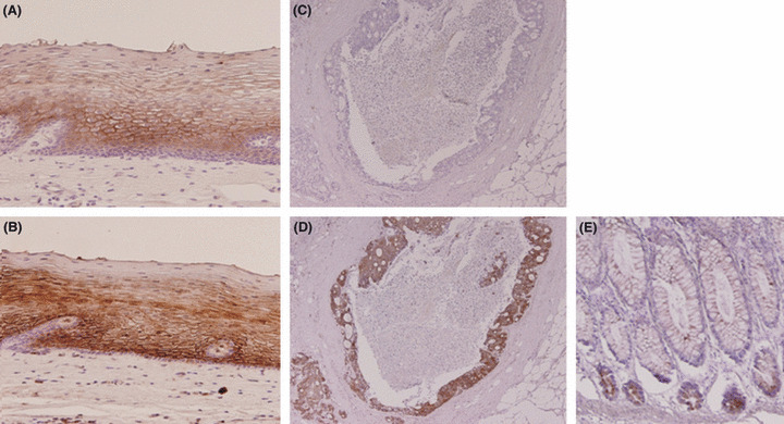
Figure 4.

Tissue sections of normal esophageal mucosa (A,B) and colorectal cancer tissue (C,D) were immunostained with anti‐C4.4A‐1 (A,C) and anti‐C4.4A‐2 (B,D) antibodies. (A,B) Membranous staining was observed in the suprabasal squamous epithelium. (C) No signal was observed with the C4.4A‐1 antibody in a colorectal cancer tissue sample. (D) Strong staining was observed with anti‐C4.4A‐2 antibody on the same sample. (E) Tissue sections of normal colonic mucosa. In normal colonic mucosa, staining was observed with anti‐C4.4A‐2 antibody at the bottom of the gland. Magnification, (A,B) ×100; (C–E) ×40.
