Figure 5.

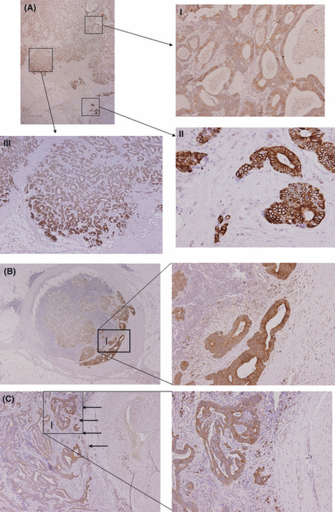
Immunohistochemistry of C4.4A expression in colorectal cancer. (A) Unique expression pattern of C4.4A at the invasive front of specimens from colorectal cancer. (A‐I) In the superficial or intermediate portion, weak C4.4A expression was observed mostly in the cytoplasm. (A‐II) C4.4A expression was present on the plasma membrane at the invasive front. (A‐III) Among the expansive population of cancer specimens, only those cells that faced virgin stroma expressed the C4.4A protein. (B) Metastatic lesion to lymph node. Metastatic tumor cells invading into the fibrous capsule exclusively expressed intense, membranous C4.4A. (C) Metastatic lesion from the lung. Metastatic tumor cells displayed a similar expression pattern of C4.4A at the invasive margin (indicated by arrows). Magnification, (A) upper left, ×12.5; (A‐I,II) ×100; (A‐III) ×40; (B) left, ×12.5; right, ×100; (C) left, ×40; right, ×100.
