
Figure 1.
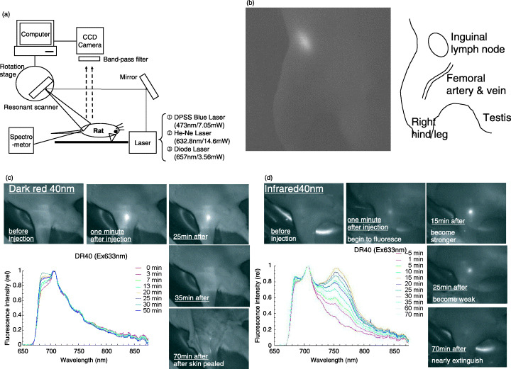
Sentinel node imaging using nano‐sized fluorescent particle in the rat. (a) Instrumentation for fluorescence imaging system. (b) Fluorescence image of right inguinal node and its illustration. (c) Fluorescence image and spectral analysis of right inguinal node after injection of dark red 40 nm‐beads. Spectral analysis shows that emission is strongest 30 min after injection. (d) Fluorescence image and spectral analysis of right inguinal node after injection of infrared 40 nm‐beads. Spectral analysis shows that emission is strongest 15 min after injection.