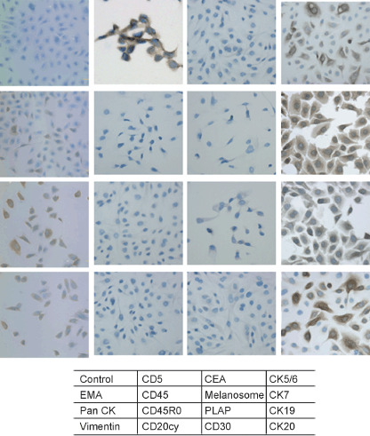
Figure 1.

Immunohistochemical staining of ThyL‐6 cells. To investigate the expression profiles of ThyL‐6 cells, the cells were attached to an eight‐chamber slide and were treated with several primary antibodies and then visualized using a HISTOFINE SAB‐PO kit according to the manufacturer's protocol. CEA: carcinoembryonal antigen; CK: cytokeratin; EMA: epithelial membrane antigen; PLAP: placental alkaline phosphatase.
