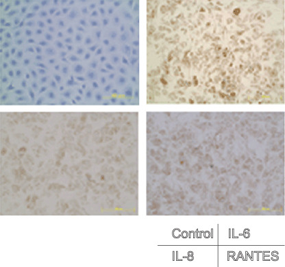
Figure 3.

Immunohistochemical staining of inflammatory cytokines. Cells attached to an eight‐chamber slide were fixed with 10% formaldehyde, and the cells were stained with antihuman interleukin (IL)‐6, IL‐8 or regulated upon activation, normal T‐cell expressed, and secreted (RANTES) antibodies, respectively, and then visualized using a HISTOFINE SAB‐PO kit.
