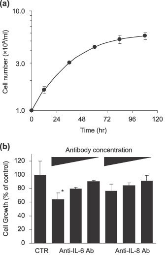
Figure 5.

(a) Cell growth curve of ThyL‐6 cells. 1 × 10⁵ cells/mL of ThyL‐6 cells were cultured in Roswell Park Memorial Institute (RPMI)‐1640 medium with 10% fetal bovine serum (FBS), and the cell growth was evaluated by the trypan‐blue dye exclusion method. Data represent the mean ± SD of triplicate cultures. (b) Growth inhibition of ThyL‐6 cells in the presence of neutralizing antibodies. Trypsinized exponential growing cells were seated on a 96‐well plate at a concentration of 1 × 10⁵ cells and incubated overnight. Following the preincubation, the cells were washed three times with PBS and resuspended in fresh medium including neutralizing interleukin (IL)‐6 or IL‐8 antibodies at a final concentration of 10%, 1%, or 0.1% of culture medium. The cells were incubated for an additional 24 h, and the growth inhibition was investigated using an MTT assay. Data represent the mean ± SD of triplicate cultures. *indicates growth rate significantly decreased (P < 0.05) in comparison with that of the untreated control cells. CTR: control; Ab: antibody.
