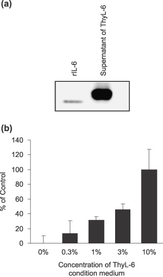
Figure 6.

(a) Western blotting detection of interleukin (IL)‐6 in the cultured supernatant of ThyL‐6 cells. The supernatant was resolved with 15% of sodium dodecylsulfate–polyacrylamide gel electrophoresis (SDS‐PAGE), and the separated proteins were transferred onto polyvinylidene difluoride membrane. The membrane was incubated with antihuman IL‐6 monoclonal antibody. Secondary antibodies conjugated to horseradish peroxidase (HRP) and a BM Chemiluminescence Western Blotting Kit were used to develop images in a Chemiluminescence Image Analyzer. (b) Cell growth of IL‐6‐dependent ILKM‐3 cells in the presence of condition medium of ThyL‐6 cells. Cells were cultured in Roswell Park Memorial Institute (RPMI)‐1640 medium with 10% fetal bovine serum (FBS) in the presence or absence of appropriate concentrations of ThyL‐6 condition medium. Cell growth was detected by an MTT assay kit. The percentage of ThyL‐6 condition medium shown represents the final concentration in the culture medium. Data represent the mean ± SD from three independent experiments.
